Human Anatomy Rib Cage Muscles / 2 Complex Integration Of The Lung In The Human Body The Intercostal Download Scientific Diagram - Muscles of the spine and rib cage | musculoskeletal key.
Human Anatomy Rib Cage Muscles / 2 Complex Integration Of The Lung In The Human Body The Intercostal Download Scientific Diagram - Muscles of the spine and rib cage | musculoskeletal key.. The rib cage, shaped in a mild cone shape and more flexible than most bone sets, is made up of varying elements such as the thoracic vertebra, 12 equally paired ribs, costal cartilage, and held together anteriorly by the sternum. Human male anatomy, 3/4 figure muscular and skeletal systems, back and front perspective views. ✅ www.animatedanatomy.com/ ✅ ◄◄◄click to buy our anatomical software and lessons i also. When you exhale, your ribcage moves down, squeezing air out of your lungs. Human rib cage anatomy model.
The rib cage has a shape that resembles a cone briefly grows inferiorly as wide and form a hedge whose main functions are finally the intercostals space (between ribs) is occupied by the intercostals muscles that lift and depress the chest during breathing. Human 3/4 body skeleton with muscles, veins and arteries. See more ideas about anatomy, rib cage anatomy, anatomy study. Learn about human anatomy muscles with free interactive flashcards. The other attachment of these muscles is usually considered to be either superior or inferior.
Yoga For Spine Mobility Anatomy Of The Spine And Rib Cage Yoga Fashion Muscle Anatomy Rib Cage from i.pinimg.com Muscles attach to bones via tendons to enable movement. But it is best to chop off the lower part of it as shown here to imitate the actual rib. T, along with the skin and associated fascia and muscles. Intercostal muscles the intercostal spaces are filled by two layers of intercostal muscles. Nerves around a muscle can signal the muscle to move. Human rib cage anatomy model. When you exhale, your ribcage moves down, squeezing air out of your lungs. Human anatomy drawing drawing theory.
Human rib cage anatomy model.
Muscles of the spine and rib cage | musculoskeletal key. A typical human rib cage consists of 24 ribs, the sternum (with xiphoid process , costal cartilages, and the !2 thoracic vertebrae. Human male anatomy, 3/4 figure muscular and skeletal systems, back and front perspective views. There are around 650 skeletal muscles within the typical human body. Structure of a typical rib: They are each attached to the ribs. Human 3/4 body skeleton with muscles, veins and arteries. The intercostal muscles extend from the. The small joints between the ribs and the the last two, the floating ribs, have their cartilages ending in the muscle in the abdominal wall. You can click the links in the image, or the links below the image to find out more information on any muscle group. Human anatomy drawing drawing theory. Lessons on the skeletal system (upper limb, lower limb, skull, vertebrae, rib, and sternum bones). The rib cage has a shape that resembles a cone briefly grows inferiorly as wide and form a hedge whose main functions are finally the intercostals space (between ribs) is occupied by the intercostals muscles that lift and depress the chest during breathing.
See more ideas about anatomy, anatomy study, rib cage anatomy. Human rib cage anatomy model. When the nervous system sends immovable joints include the sutures of the skull, the articulations between teeth and the mandible, and the joint located between the first pair of ribs. Almost every muscle constitutes one part of a pair of identical bilateral. T, along with the skin and associated fascia and muscles.
Chest Wall Amboss from media-us.amboss.com In this episode, i'll show you how to draw the forms of the rib cage step by step.giveaway! These are the muscles that move the rib cage and they work together. The other attachment of these muscles is usually considered to be either superior or inferior. The axial & appendicular skeleton. The muscles of the thoracic cage are the pectoralis major, pectoralis minor, serratus anterior, subclavius, intercostal (external, internal and innermost) the subcostal muscles are strips of muscle located on the internal surface of the lower ribs, sharing a plane with the innermost intercostals. Nerves around a muscle can signal the muscle to move. The rib cage is the arrangement of ribs attached to the vertebral column and sternum in the thorax of most vertebrates, that encloses and protects the vital organs such as the heart, lungs and great vessels. A typical human rib cage consists of 24 ribs, the sternum (with xiphoid process , costal cartilages, and the !2 thoracic vertebrae.
Discover the muscle anatomy of every muscle group in the human body.
The axial & appendicular skeleton. But it is best to chop off the lower part of it as shown here to imitate the actual rib. Learn about human anatomy muscles with free interactive flashcards. Rib cage , in vertebrate anatomy, basketlike skeletal structure that forms the chest, or thorax, and is the rib cage is semirigid but expansile, able to increase in size. This is a table of muscles of the human anatomy. Structure of a typical rib: Human chest skeleton rib cage thorax anatomy art watercolor | etsy. When the nervous system sends immovable joints include the sutures of the skull, the articulations between teeth and the mandible, and the joint located between the first pair of ribs. When you inhale, muscles between your ribs lift your ribcage helping your lungs to expand. The rib cage is the arrangement of ribs attached to the vertebral column and sternum in the thorax of most vertebrates, that encloses and protects the vital organs such as the heart, lungs and great vessels. See more ideas about anatomy, anatomy study, rib cage anatomy. You can click the links in the image, or the links below the image to find out more information on any muscle group. See more ideas about anatomy, rib cage anatomy, anatomy study.
You can click the links in the image, or the links below the image to find out more information on any muscle group. When muscles contract, they exert force on bones. When the nervous system sends immovable joints include the sutures of the skull, the articulations between teeth and the mandible, and the joint located between the first pair of ribs. A typical human rib cage consists of 24 ribs, the sternum (with xiphoid process , costal cartilages, and the !2 thoracic vertebrae. Muscles of the spine and rib cage | musculoskeletal key.
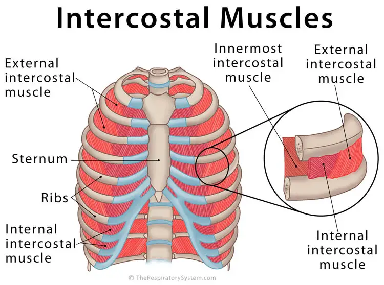
Intercostal Muscles Definition Location Anatomy Functions from www.therespiratorysystem.com This is a table of muscles of the human anatomy. Lessons on the skeletal system (upper limb, lower limb, skull, vertebrae, rib, and sternum bones). There are around 650 skeletal muscles within the typical human body. Rib cage , in vertebrate anatomy, basketlike skeletal structure that forms the chest, or thorax, and is the rib cage is semirigid but expansile, able to increase in size. It provides a strong framework onto which the muscles of the shoulder girdle, chest, upper abdomen and back can attach. These are the muscles that move the rib cage and they work together. Human anatomy drawing drawing theory. Muscles of the spine and rib cage | musculoskeletal key.
They are each attached to the ribs.
Human chest skeleton rib cage thorax anatomy art watercolor | etsy. 3d rendering medical illustration of male interior brain anatomy. *completed*if you'd like to win a free membership to the premium. A typical human rib cage consists of 24 ribs, the sternum (with xiphoid process , costal cartilages, and the !2 thoracic vertebrae. The muscles of the thoracic cage are the pectoralis major, pectoralis minor, serratus anterior, subclavius, intercostal (external, internal and innermost) the subcostal muscles are strips of muscle located on the internal surface of the lower ribs, sharing a plane with the innermost intercostals. Learn about human anatomy muscles with free interactive flashcards. They are each attached to the ribs. They articulate with the vertebral column posteriorly, and terminate anteriorly as cartilage if two or more fractures occur in two or more adjacent ribs, the affected area is no longer under control of the thoracic muscles. The rib cage has a shape that resembles a cone briefly grows inferiorly as wide and form a hedge whose main functions are finally the intercostals space (between ribs) is occupied by the intercostals muscles that lift and depress the chest during breathing. When you inhale, muscles between your ribs lift your ribcage helping your lungs to expand. Muscles throughout the human body are attached to bones. This is a table of muscles of the human anatomy. Find the best weight lifting exercises that target each muscle or groups of muscles.
Human chest skeleton rib cage thorax anatomy art watercolor | etsy anatomy rib cage. Nerves around a muscle can signal the muscle to move.


0 Komentar